
Why Many Eczema Treatments Provide Relief — But Not Lasting Resolution
And What It Means to Treat Atopic Dermatitis as a Multifactorial Disease
Author: Dr. Rafal Pielak
Atopic dermatitis (AD) is the most common inflammatory skin disease worldwide, affecting an estimated 15–20% of children and up to 10% of adults. (Langan et al., 2020) For millions of people, it is far more than a rash—it is a chronic condition defined by relentless itch, disrupted sleep, compromised skin integrity, and a significant psychosocial burden. Despite a remarkable expansion of therapeutic options over the past decade, a fundamental challenge remains: most treatments effectively suppress symptoms, yet few address the underlying biological processes that allow the disease to develop and persist.
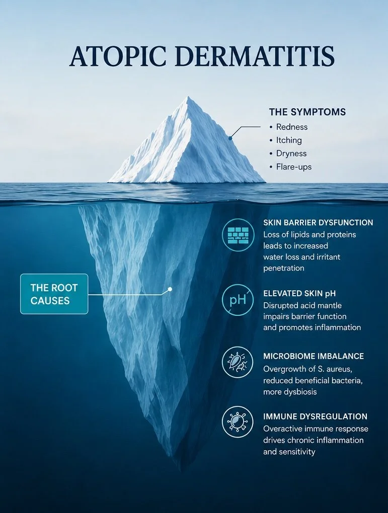
Atopic dermatitis is not driven by a single pathway, but rather represents a multifactorial, systems-level disorder (Elias et al. 2012, Elias & Wakefield, 2014). It emerges from the interplay of several interconnected components: skin barrier dysfunction, disruption of the acid mantle and elevation of surface pH, microbiome imbalance, and immune dysregulation. Importantly, the immune response, while central to disease expression, is often a downstream consequence rather than the initiating event. Increasing evidence points to the earliest trigger as a disturbance of the skin barrier and acid mantle, accompanied by a shift toward elevated surface pH (Schmid-Wendtner & Korting, 2006, Pielak & Maibach, 2022). This initial disruption sets off a cascade of biochemical and microbiological changes that ultimately amplify inflammation and chronicity.
In this article, we explore the full biological landscape of atopic dermatitis through the lens of its core disease pillars. We examine why most current therapies focus narrowly on a single dimension—primarily immune modulation—and consider what a more integrative approach might achieve. By combining immune control with restoration of the skin’s acid mantle and pH homeostasis, it may be possible not only to manage symptoms more effectively, but to intervene closer to the root of the disease and improve outcomes across all stages of severity.
The Standard Approach: Targeting Inflammation
The range of treatments available for eczema today is, by any measure, impressive. Over the past two decades, the field has advanced from almost total reliance on broad steroid creams to a growing menu of highly targeted therapies. Understanding what these treatments do — and what they don't — is the first step toward seeing why a more complete approach is needed.
Topical Corticosteroids (TCS)
Still the backbone of acute flare management, topical corticosteroids work by broadly suppressing the skin’s inflammatory response. (Wollenberg et al., 2020) They reduce the production of pro-inflammatory cytokines, dampen immune cell activity in the dermis, and quickly relieve itch and redness. After more than 60 years of use, their effectiveness is well established. However, they do not address the underlying drivers of the disease—skin barrier dysfunction, disruption of the acid mantle, or microbiome imbalance. And with long-term use, they carry well-documented risks, including skin atrophy, striae, telangiectasias, and, with more potent formulations, the potential for systemic absorption.
Topical Calcineurin Inhibitors (TCIs): Tacrolimus and Pimecrolimus
These agents inhibit calcineurin-dependent T-cell activation, blocking the production of key inflammatory cytokines including IL-2, IL-4, and IL-13. (Reitamo et al. 2004) They are non-steroidal and therefore avoid the atrophy risk associated with corticosteroids, making them suitable for sensitive areas like the face and skin folds. Like TCS, however, they function as immune modulators without addressing barrier function or the acid environment of the skin surface.
Phosphodiesterase-4 (PDE4) Inhibitors: Crisaborole
Crisaborole (Eucrisa) is a topical small molecule that inhibits PDE4, an enzyme that degrades cyclic AMP. (Hanifin et al. 2016) By elevating intracellular cAMP levels, it dampens inflammatory signaling across multiple immune cell types. It is effective in mild-to-moderate disease and well-tolerated, but again operates entirely within the immune compartment.
Biologics: Dupilumab, Tralokinumab, Lebrikizumab, Nemolizumab
The introduction of dupilumab (Dupixent) in 2017 represented a genuine inflection point. By blocking the shared receptor subunit for IL-4 and IL-13 (the two cytokines most central to the Th2-skewed immune response in AD) dupilumab produces dramatic and durable responses in moderate-to-severe disease. (Simpson et al. 2016) Tralokinumab and lebrikizumab target IL-13 specifically with comparable efficacy. (Silverberg et al. 2021) More recently, nemolizumab has emerged as an exciting option targeting IL-31 receptor alpha, directly addressing the pruritic signaling axis. (Ruzicka et al. 2017) These biologics represent the closest thing the field has to disease-modifying therapy. And yet they, too, act exclusively on the immune and neuroimmune axis.
JAK Inhibitors: Upadacitinib, Abrocitinib, Ruxolitinib
Janus kinase inhibitors block intracellular signaling downstream of multiple cytokine receptors simultaneously. Oral JAK inhibitors such as upadacitinib (Rinvoq) and abrocitinib (Cibinqo) are among the most potent therapies available for AD, with rapid onset and high EASI responder rates in clinical trials. Topical ruxolitinib (Opzelura) offers a localized option with a favorable safety profile. As with all the therapies above, their mechanism of action is immunological. They do not address the skin surface chemistry that sets the stage for disease.
“Every approved pharmacotherapy for atopic dermatitis acts on the immune system. Not one is designed to restore the acid mantle, repair the physical barrier, and rebalance the skin microbiome.”
Atopic Dermatitis Is Not Just an Immune Disease
To understand why immune-only treatments can only go so far, we need to map the full terrain of atopic dermatitis pathophysiology. AD is best understood not as a single disease but as a convergence of several interconnected biological failures, each capable of driving the others in a self-reinforcing cycle. (Paller et al. 2019)
ㅤ1. Disrupted Acid Skin Acid Mantle and Elevated pH
Healthy skin maintains a slightly acidic surface pH of ~4.7. (Lambers et al. 2006, Pielak & Maibach 2022) This acidic environment, known as the acid mantle, is not incidental. It is the master regulator of virtually every aspect of skin function: enzyme activity, barrier lipid organization, microbial ecology, and immune signaling at the skin surface. (Elias 2005)
The acid mantle is generated primarily through three mechanisms: the secretion of organic acids (lactic acid, amino acids, and pyrrolidone carboxylic acid) from eccrine glands and the natural moisturizing factor pool; the hydrolysis of phospholipids in the stratum corneum, which liberates free fatty acids (Fluhr et al. 2004); and the acidifying activity of profilaggrin-derived metabolites and V-type H⁺-ATPases embedded in lamellar body membranes. (Elias 2005) Together, these mechanisms create and maintain an environment inhospitable to pathogenic bacteria, optimized for barrier enzyme activity, and calibrated to suppress aberrant immune activation at the skin surface.
In atopic dermatitis, this pH is dramatically elevated — often to values between 5.5 and 6.5, sometimes approaching neutrality. (Hatano et al. 2009) This is not a trivial perturbation. A change of just 0.5–1.0 pH units shifts enzymatic kinetics, alters membrane protein conformation, and fundamentally changes what can survive on the skin surface. The elevated pH in AD is both a consequence and an amplifier of disease, and it sits at the top of the pathophysiological cascade.
ㅤ2. Leaky Barrier: Structural Skin Failureㅤ
The skin barrier is a sophisticated multilayer defense organized in a mortar-and-brick architecture in which corneocytes (the bricks) are embedded in a matrix of ceramides, free fatty acids, and cholesterol (the mortar). This lipid lamellar matrix is not merely structural; it is the primary physical impediment to transepidermal water loss (TEWL) and the first line of defense against environmental allergens, irritants, and microbial pathogens. (Proksch et al. 2008)
In atopic dermatitis, this barrier is profoundly compromised. Loss-of-function mutations in the filaggrin gene (FLG) — present in approximately 30–40% of AD patients — reduce the production of filaggrin, a structural protein critical for corneocyte maturation and the generation of natural moisturizing factor (NMF). (Palmer et al. 2006; Sandilands et al. 2009) NMF components include urocanic acid, which contributes to the skin's pH buffering capacity, and pyrrolidone carboxylic acid, a potent humectant. Without adequate filaggrin and NMF, the corneocyte scaffold collapses, the stratum corneum becomes disorganized, and water escapes freely from the epidermis.
Crucially, the enzymes responsible for processing the precursor lipids that form the lamellar membrane, such as serine ceramidases, beta-glucocerebrosidase, and acidic sphingomyelinase, are pH-sensitive. They require an acidic environment to function optimally. (Elias & Schmuth 2009; Vávrová et al. 2014) When skin pH rises, these enzymes are inhibited, ceramide synthesis is impaired, and the mortar between the bricks is literally dissolved. (Choi et al. 2024) The barrier breaks not only because of genetic predisposition but because the chemical environment needed to build and maintain it has been destroyed.
ㅤ3. Immune Dysregulation: Consequence, Not Cause
The Th2-dominant immune phenotype of atopic dermatitis, characterized by elevated IL-4, IL-13, IL-31, IL-33, and TSLP, is well established and clinically significant. (Brandt & Sivaprasad 2011) These cytokines drive IgE production, mast cell activation, eosinophil recruitment, and the suppression of the Th1 response needed for antimicrobial defense. They also directly suppress the expression of filaggrin and other barrier proteins, further destabilizing the skin structure. (Kim et al. 2008)
What is less often discussed is the directionality of this immune activation. Elevated skin surface pH and barrier breach create the conditions for immune alarm signals. Epithelial-derived cytokines, particularly TSLP (thymic stromal lymphopoietin) and IL-33, are released in response to barrier disruption and allergen penetration. (Soumelis et al. 2002) These alarmins prime dendritic cells and innate lymphoid cells (ILC2s) toward a Th2 phenotype. In other words, the barrier failure and pH disturbance come first; the immune response follows. This does not mean immune-directed therapy is misguided — by the time a patient reaches a dermatologist, the immune response is well established and must be addressed. But it does mean that treating only the immune response leaves the original insult unresolved. (Leung & Guttman-Yassky 2014)
ㅤ4. Microbiome Imbalance: The Microbial Dimension
The skin surface is home to a rich and diverse community of microorganisms — bacteria, fungi, and viruses — collectively known as the cutaneous microbiome. In healthy skin, this community is dominated by beneficial commensals such as Staphylococcus epidermidis, which actively suppress pathogen colonization, stimulate antimicrobial peptide production, and maintain a stable, mildly acidic surface ecology. (Gallo & Nakatsuji 2011; Nakatsuji et al. 2017)
In atopic dermatitis, the skin microbiome undergoes a dramatic and well-characterized shift. During flares, Staphylococcus aureus can constitute more than 90% of the bacterial community on affected skin — displacing the beneficial commensals that would otherwise limit its proliferation. (Totté et al. 2016) S. aureus is not a passive bystander. It secretes a battery of virulence factors — including proteases (V8 protease, aureolysin), toxins (alpha-toxin, delta-toxin, superantigens), and sphingomyelinases — that directly damage the skin barrier, activate keratinocyte-derived alarmins, and amplify the Th2 immune response. (Brüssow 2020) Its superantigens stimulate T-cell activation independently of antigen presentation, maintaining a state of chronic immune activation even in the absence of allergens. (Cardona et al. 2006, Kim et al. 2006)
The relationship between S. aureus and skin pH is direct and mechanistic. S. aureus grows optimally at pH 7.0–7.5 and is inhibited at pH below 5.5. (Wollenberg & Wetzel 2003) The elevated pH of AD skin is therefore not merely a marker of disease — it is a permissive environment that allows S. aureus to establish dominance and sustain itself. This is why restoring skin acidity is not just a cosmetic consideration but a direct antimicrobial intervention.
ㅤ5. Neurosensory Sensitivity: The Itch–Scratch Cycle
Chronic pruritus — itch — is arguably the most debilitating feature of atopic dermatitis and the one least well addressed by conventional anti-inflammatory therapy. (Yosipovitch & Bernhard 2013) Itch in AD is mediated through a complex network of peripheral sensory neurons, epithelial cells, and immune mediators. Elevated IL-31, released by Th2 cells and mast cells, directly activates IL-31 receptor alpha on sensory nerve fibers, triggering itch signals that bypass the histamine pathway (Cevikbas et al. 2014) — which is why antihistamines are largely ineffective in AD. TSLP, periostin, and substance P further sensitize sensory neurons, lowering the threshold for itch perception. (Bautista et al. 2015)
The scratch response to itch perpetuates barrier damage, introduces S. aureus from under the fingernails, and triggers further release of inflammatory mediators. This itch–scratch cycle is a self-sustaining loop that can maintain disease activity even after other triggers have been removed. (Bautista et al. 2015) Neurosensory sensitization in AD is also linked to small fiber neuropathy in affected skin regions — a finding that suggests structural changes in the peripheral nervous system as a consequence of chronic inflammation — changes that do not reverse quickly even when inflammation is controlled. (Tominaga & Takamori 2014)
The pH Link: A Master Switch Connecting All Five Pillars
If atopic dermatitis is a systems disease involving five interconnected failures, then any truly effective intervention must address multiple nodes in the system simultaneously. And when we look carefully at the biology, one variable sits at the intersection of every pillar: skin surface pH.
Acid mantle disruption does not merely coexist with barrier dysfunction, immune dysregulation, microbiome imbalance, and neurosensory sensitization. It is mechanistically intertwined with each of them. The consequences of elevated skin pH propagate across multiple interconnected biological systems involved in atopic dermatitis pathogenesis:
Elevated pH dysregulates epidermal protease activity. Acidification normally restrains serine proteases such as kallikrein-5 (KLK5) and kallikrein-7 (KLK7), which are required for controlled desquamation. When pH rises, this regulation breaks down, resulting in excessive protease activity, accelerated corneocyte shedding, and progressive thinning of the stratum corneum barrier. (Hachem et al. 2006)
Elevated pH impairs lipid-processing enzymes essential for barrier formation. Key enzymes involved in generating the lamellar lipid matrix, including secretory phospholipase A2, β-glucocerebrosidase, and acidic sphingomyelinase, require an acidic environment for optimal function. Increased pH reduces ceramide production and disrupts assembly of the extracellular lipid bilayer, directly compromising barrier integrity. (Vávrová et al. 2014)
Elevated pH disrupts the structural organization of lipid lamellae. Beyond enzyme activity, acidic conditions help maintain the electrostatic interactions that organize lipid membranes within the extracellular space. pH elevation contributes to lamellar body extrusion defects and disorganization of the barrier architecture, even when lipid precursors remain present. (Elias 2005)
Elevated pH reshapes the skin microbiome toward dysbiosis. A more alkaline environment favors the growth and virulence of Staphylococcus aureus while suppressing beneficial commensal organisms such as Staphylococcus epidermidis. These commensals normally produce antimicrobial peptides, short-chain fatty acids, and phenol-soluble modulins that help suppress pathogen colonization and maintain immune balance. (Kobayashi et al. 2015; Nakatsuji et al. 2017)
Elevated pH facilitates immune activation and allergic sensitization. Barrier disruption permits increased penetration of environmental allergens, microbial toxins, and protease signals into the viable epidermis. This stimulates keratinocyte-derived cytokines such as TSLP and IL-33, which initiate and amplify downstream Th2-skewed immune responses characteristic of atopic dermatitis. (Nakatsuji et al. 2016; Soumelis et al. 2002)
Elevated pH may contribute directly to itch sensitization. Altered extracellular pH can modulate pH-sensitive ion channels, including ASICs (acid-sensing ion channels) and TRPV1 on cutaneous sensory neurons, potentially lowering the threshold for itch perception and contributing to chronic neurosensory hypersensitivity. (Cevikbas et al. 2014)
“Restoring physiological skin pH is not one intervention among many — it is an upstream intervention that simultaneously addresses barrier repair, microbial ecology, immune tone, and potentially neurosensory threshold.”
This is the promise of acid mantle restoration as a therapeutic strategy. By reacidifying the skin surface to the physiological range of pH 4.7–5.0, it is possible to re-enable the enzymes that build and maintain the barrier, recreate the chemical environment that favors commensals over pathogens, reduce the penetration of immune-activating signals, and quiet the chronic low-grade alarm state that perpetuates the disease.
An Integrative Framework: Matching Treatment Depth to Disease Severity
The recognition that AD is a multifactorial systems disease — and that pH restoration addresses multiple disease nodes simultaneously — suggests a stratified therapeutic framework in which the choice of intervention is matched to the severity and complexity of the disease state.
Mild-to-Moderate AD: Acid Mantle Restoration as Standalone Therapy
For patients with mild-to-moderate atopic dermatitis — those with limited body surface area involvement, manageable itch, and preserved quality of life — the evidence increasingly supports that targeted restoration of the skin's acid mantle may be sufficient as a primary therapeutic intervention, provided it is delivered with the precision and formulation science that the biology demands. (Elias et al. 2012)
Effective acid mantle restoration requires intentional control of skin surface pH through formulations designed to operate within the physiological range of healthy skin, ideally around pH 4.5–5.0. Equally critical is the ability to maintain that acidic environment over time through adequate buffering capacity and the use of ingredients that support the skin’s endogenous acidification mechanisms, rather than temporarily lowering pH with a transient acidic preparation.
If this physiological environment can be effectively restored and maintained, several downstream effects may follow. Reacidification may help normalize kallikrein protease activity, improving desquamation dynamics and reducing excessive protease-mediated barrier disruption. (Hachem et al. 2006) Lipid-processing enzymes may recover function, potentially supporting ceramide synthesis and reorganization of the extracellular lipid matrix. (Vávrová et al. 2014) At the same time, a more acidic skin surface may shift the microbial ecosystem away from Staphylococcus aureus dominance and toward a healthier commensal balance. (Nakatsuji et al. 2017)
Through these interconnected mechanisms, restoration of the acid mantle could potentially reduce transepidermal water loss, decrease penetration of allergens and microbial products, and dampen chronic immune activation arising from a barrier-compromised, dysbiotic skin surface. Rather than acting primarily through direct immunosuppression, this strategy proposes that improving the underlying biochemical environment of the skin may allow multiple pathogenic pathways to gradually move back toward homeostasis. In this framework, acid mantle restoration is not positioned as a replacement for all existing therapies, but as a systems-level approach that may offer meaningful clinical benefit, particularly in earlier or less severe disease states.
Moderate-to-Severe AD: Combination Therapy — Immunomodulation Plus Acid Mantle Restoration
n moderate-to-severe atopic dermatitis, the disease may progress beyond a state that can be adequately controlled through restoration of skin surface chemistry alone. At this stage, immune dysregulation is often deeply established, with cytokine-driven suppression of barrier function actively contributing to disease persistence. IL-4 and IL-13, for example, have been shown to downregulate key structural proteins involved in epidermal barrier formation, including filaggrin, loricrin, and involucrin. (Kim et al. 2008; Bao et al. 2017) At the same time, Staphylococcus aureus colonization may become more entrenched, while chronic itch signaling and neurosensory sensitization further reinforce the inflammatory cycle.
Within this context, immunomodulatory therapies such as biologics and JAK inhibitors may play an essential role in reducing the inflammatory burden and interrupting cytokine-driven disease amplification. However, a systems-level framework suggests that immunomodulation and acid mantle restoration should not necessarily be viewed as competing strategies, but rather as potentially complementary interventions acting on different components of the same interconnected disease network.
In this proposed model, biologics and JAK inhibitors primarily target inflammatory signaling pathways that suppress barrier gene expression and perpetuate itch and immune activation. Acid mantle restoration, by contrast, may help address parallel processes occurring at the skin surface, including elevated pH, impaired lipid-processing enzyme activity, microbiome dysbiosis, and increased permeability to allergens and microbial products. Together, these approaches could theoretically influence multiple layers of disease pathophysiology simultaneously rather than focusing on a single pathway in isolation.
This combinatorial rationale is supported by mechanistic considerations. If cytokine signaling is reduced through biologic therapy, but the skin surface environment remains alkaline and dysbiotic, recovery of barrier function may still be limited because the enzymatic machinery required for proper lipid processing and barrier assembly continues to operate under unfavorable biochemical conditions. (De Benedetto et al. 2011) Conversely, attempts to restore the acid mantle alone in severe disease may be insufficient if persistent cytokine activity continues to suppress production of critical barrier proteins.
A systems-level therapeutic strategy therefore proposes that simultaneous modulation of both immune signaling and skin surface physiology may produce synergistic effects. By reducing inflammatory suppression from within while improving the biochemical environment at the skin surface, each intervention may help remove constraints that limit the effectiveness of the other.
A Vision for the Future: Acid Mantle Restoration as a Biologic-Sparing Strategy
Perhaps the most clinically exciting implication of an integrative acid mantle approach concerns the long-term trajectory of moderate-to-severe AD treatment — specifically, the possibility that systematic barrier restoration combined with immunomodulation could, over time, reduce or eventually eliminate the need for ongoing biologic therapy.
This is not an implausible aspiration. It is grounded in the following mechanistic reasoning.
Biologics Suppress the Consequence; Barrier Restoration Removes the Cause
Dupilumab, tralokinumab, and their counterparts are extraordinarily effective at controlling the Th2 cytokine milieu that characterizes active AD. But they do not cure the disease — when treatment is discontinued, the majority of patients relapse. (Abuabara et al. 2022) This is because the underlying conditions that generate the immune stimulus — the leaky, alkaline, S. aureus-colonized skin surface — remain unresolved. The immune system responds to signals coming from the skin; if those signals persist, the Th2 response will resume once the pharmacological brake is lifted. (Elias & Wakefield 2011)
Acid mantle restoration, deployed alongside biologic therapy, addresses the source of those signals. By restoring the barrier, rebalancing the microbiome, and reducing the continuous influx of immune-activating stimuli from the skin surface, it gradually diminishes the biological pressure that drives the Th2 response. (Nakatsuji et al. 2016) The immune system, no longer being continuously alarmed by a breached, alkaline, dysbiotic skin surface, can shift back toward homeostasis.
A Possible Tapering Framework
In clinical practice, this would argue for an induction phase in which a biologic is initiated at standard dosing to rapidly control active inflammation, (Simpson et al. 2016) while concurrent acid mantle restoration begins the slower, surface-level work of repairing the barrier and rebalancing the microbiome. Over 6 to 12 months, as barrier integrity improves and S. aureus colonization declines, the inflammatory stimulus diminishes. (Guttman-Yassky et al. 2019) At this point, it becomes clinically rational to explore careful, supervised tapering of the biologic — extending injection intervals, for example, from every two weeks to every four weeks — while monitoring closely for evidence of flare.
For a subset of patients — likely those with mild-to-moderate underlying disease who were pushed into the moderate-to-severe category by an unaddressed pH and barrier deficit — full discontinuation of biologic therapy may become achievable. For others with more severe intrinsic disease, the outcome may be a lower effective biologic dose or longer dosing interval rather than full withdrawal. Either outcome represents a meaningful improvement: reduced drug exposure, reduced cost, reduced injection burden, and a more physiological disease state.
Implications for Disease Modification
The significance of this framework extends beyond individual treatment optimization. If the acid mantle is genuinely the upstream hub from which barrier failure, immune dysregulation, and microbiome imbalance radiate, then systematic restoration of the acid mantle — initiated early, maintained consistently, and paired with immune control when needed — constitutes the closest thing we currently have to a disease-modifying strategy in atopic dermatitis. (Elias & Wakefield 2014)
Disease modification — changing the natural history of a condition rather than merely managing its symptoms — is the gold standard of therapeutic achievement in chronic inflammatory disease. It has been demonstrated in rheumatoid arthritis (where early methotrexate use can prevent joint destruction), in multiple sclerosis (where early high-efficacy therapy preserves neurological function), and increasingly in allergic disease (where sublingual immunotherapy modifies the underlying allergic response). In AD, the equivalent would be a therapeutic approach that reduces relapse rates, extends remission duration, and progressively lowers disease severity over time — not merely one that controls active flares.
An integrative strategy combining acid mantle restoration with appropriate immunomodulation offers a scientifically coherent path toward that goal. The acid mantle is not the only biological target worth pursuing in AD — but it may be the one most consequentially neglected.
What This Means for Patients and Clinicians
The practical implications of this framework are significant and actionable at every point along the severity spectrum.
For patients with mild-to-moderate AD, the message is that the products they apply to their skin are not merely moisturizers — they are therapeutic agents whose pH, formulation, and mechanism of action matter enormously. Choosing skin care products specifically formulated to restore and maintain physiological pH is not a cosmetic decision; it is a clinical one. And the expectation from such products, when they are well-designed, should not be merely comfort — it should be measurable improvement in TEWL, microbiome composition, and disease severity scores.
For patients with moderate-to-severe AD on biologic therapy, the message is that their medication is working on one node of a multi-node system. Adding precision acid mantle restoration to their regimen is not redundant — it is complementary, potentially synergistic, and opens the door to a future in which their biologic dose or frequency is reduced as their skin biology stabilizes.
For dermatologists and allergists, the framework argues for expanding the treatment conversation beyond the prescription pad. Asking about a patient's skin care routine, their product formulations, and the pH of the products they use is not trivial — it is mechanistically relevant clinical questioning. And recommending or co-prescribing acid mantle restoration products alongside systemic therapy, with the explicit goal of barrier consolidation and microbiome normalization, is a scientifically grounded intervention.
For researchers, the framework generates specific and testable hypotheses: Does systematic pH restoration reduce the biologic dose required to maintain EASI-75 response? Do patients on combination therapy show different patterns of microbiome recovery than those on biologics alone? Can acid mantle restoration initiated in infancy — in filaggrin-haploinsufficient children at high risk of AD — prevent disease development entirely? These are answerable questions with the research designs that exist today.
Conclusion: The Root Cause Is Upstream
Atopic dermatitis is not a mystery. Its biology is increasingly well understood, and the five pillars of the disease — acid mantle disruption, barrier failure, immune dysregulation, microbiome imbalance, and neurosensory sensitization — map onto each other with a coherence that points clearly toward the acid mantle as the upstream regulatory hub.
The treatments that currently dominate the AD therapeutic landscape are genuinely effective at what they do. Biologics, JAK inhibitors, and topical immunomodulators represent real advances in the management of a disease that was, until recently, largely confined to corticosteroids and emollients. But they address the disease from the middle of the cascade, not the beginning. They are powerful downstream interventions in a disease whose root cause lies upstream.
Restoring the acid mantle is the upstream intervention. It re-enables the enzymatic machinery that builds the barrier. It recreates the ecological conditions that favor commensals over pathogens. It reduces the flux of immune-activating signals from the skin surface. And deployed alongside appropriate immunomodulation in more severe disease, it creates the conditions in which the immune system no longer needs to be pharmacologically restrained — because the signals driving it have been addressed at their source.
“Treating eczema without restoring the acid mantle is like mopping the floor without turning off the tap. The immune system is responding to something. That something is coming from the skin. Fix the skin, and the immune system — often — will follow.”
The goal of atopic dermatitis management should not merely be symptom control. It should be restoration of skin biology to a state of sustained, self-maintaining health — a state in which the skin's own regulatory mechanisms, once re-enabled by physiological pH and a repaired barrier, can do what they evolved to do: protect, regulate, and defend. That is what a systems-level approach to atopic dermatitis makes possible. And that is the standard to which the field should aspire.
References
Brandt EB, Sivaprasad U. Th2 cytokines and atopic dermatitis. J Clin Cell Immunol. 2011; 2(3):110
Langan SM, Irvine AD, Weidinger S. Atopic dermatitis. Lancet. 2020; 396(10247):345–360.
Moniaga CS, Jeong SK, Egawa G, et al. Flaky tail mouse denotes human atopic dermatitis in the steady state and by sequential immunization. Am J Pathol. 2010; 176(5):2385–2393.
Yosipovitch G, Bernhard JD. Chronic pruritus. N Engl J Med. 2013; 368(17):1625–1634.