
The Acid Mantle and Epidermal Proteases: What Happens When pH Rises in Atopic Dermatitis
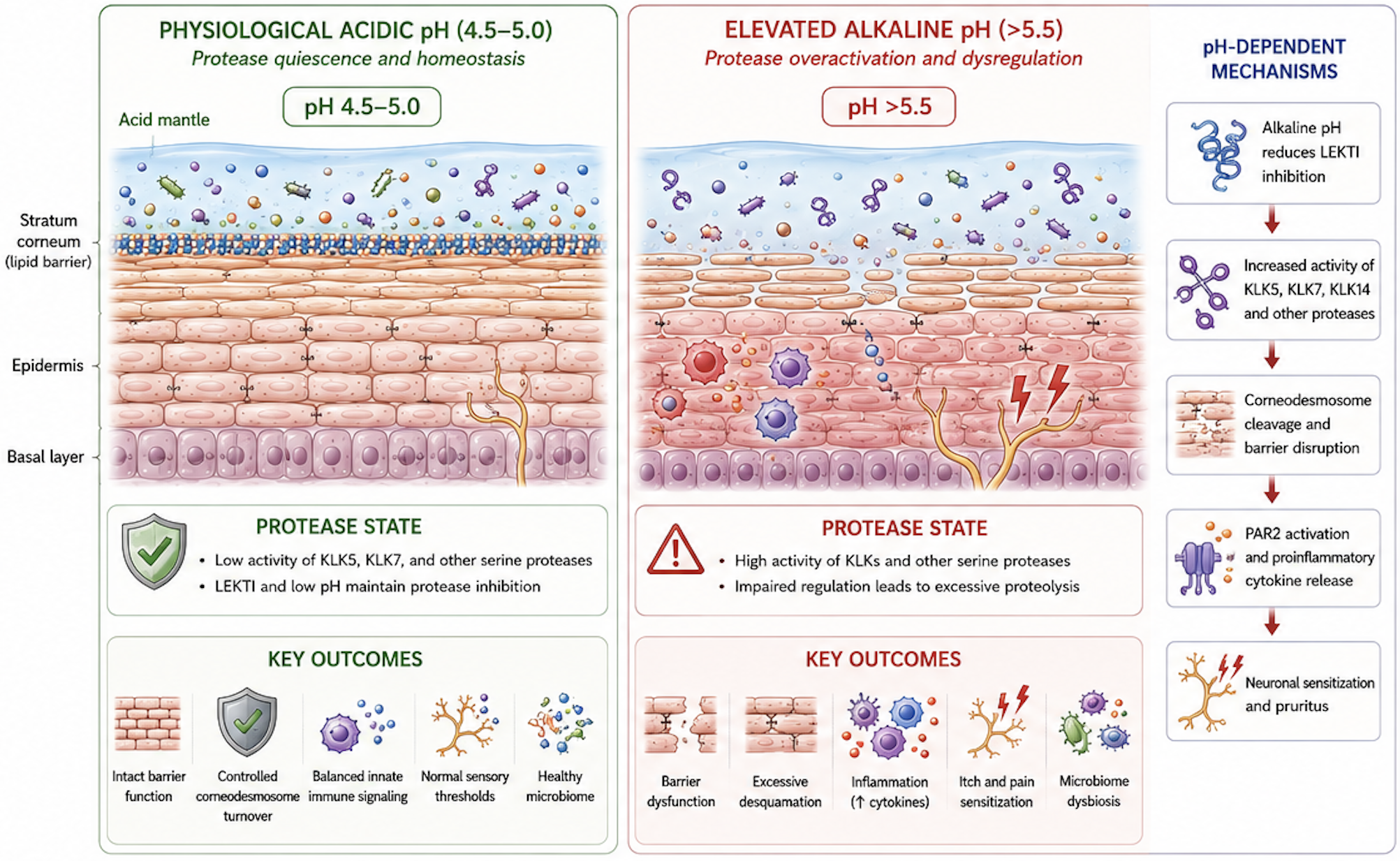
The acid mantle is often described as a thin, protective acidic film on the skin surface. That description is broadly correct, but incomplete. The acid mantle is a dynamic, continuously regenerated chemical environment that helps regulate enzymes within the stratum corneum. Among the most important are proteases that coordinate normal shedding but, when insufficiently controlled, can weaken the epidermal barrier.
This relationship is especially important in atopic dermatitis (AD), where elevated skin surface pH is a consistent finding and can contribute directly to barrier disruption, inflammation, and itch.
Author: Dr. Rafal Pielak
From Protective Film to Active Regulatory System
The concept of the acid mantle dates back nearly a century. In 1928, Schade and Marchionini observed that healthy skin is acidic relative to the near-neutral pH of internal tissues (Schade and Marchionini 1928). They called this surface environment the Säureschutzmantel (literally, the “acid protective mantle”) and it was initially understood mainly as a chemical defense against microorganisms.
Modern epidermal biology has substantially expanded that view. Stratum corneum acidity is generated and maintained through a distributed network of processes, including the production of filaggrin-derived acids, the release of free fatty acids from phospholipids and sebum, lactic acid from sweat and resident microorganisms, and localized proton transport near the base of the stratum corneum (Fluhr and Elias 2002; Elias 2015). No single pathway acts alone. Together, these processes establish and continually restore an acidic gradient toward the skin surface (Surber et al. 2018; Brooks et al. 2025).
This acidity is biologically important because many processes in the outer epidermis—including lipid processing, microbial selection, immune signaling, and enzyme activity—depend on pH.
The Enzymes That Hold the Stratum Corneum Together—and Take It Apart
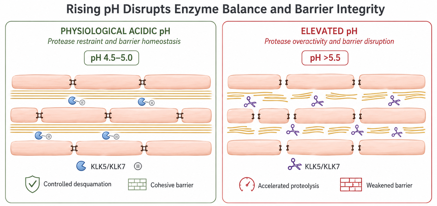
Corneocytes in the stratum corneum are connected by specialized protein structures called corneodesmosomes. For normal desquamation to occur, these structures must be progressively degraded as corneocytes move toward the surface. This process is mediated largely by serine proteases of the kallikrein family, particularly KLK5 and KLK7, which cleave corneodesmosomal proteins (Caubet et al. 2004).
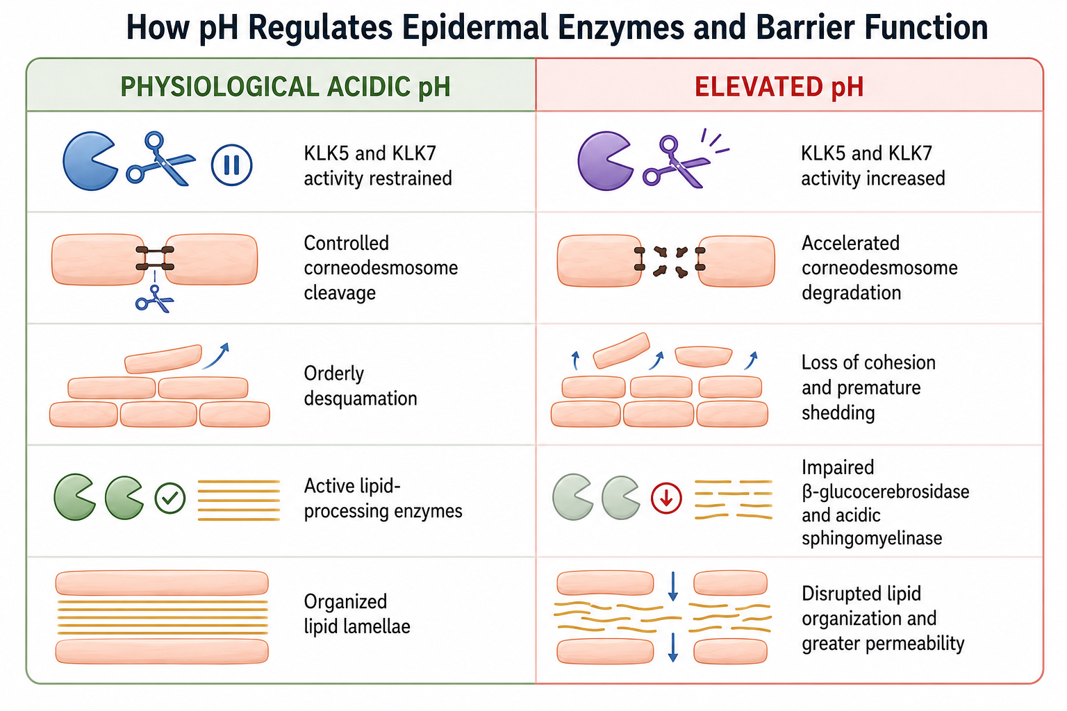
A critical feature of these enzymes is their intrinsic sensitivity to pH. Biochemical studies show that KLK5 and KLK7 are substantially more active at neutral to slightly alkaline pH than under the mildly acidic conditions normally present in the stratum corneum (Caubet et al. 2004; Brattsand et al. 2005). Acidity therefore acts as a direct biochemical restraint on the rate at which these enzymes cleave their substrates.
At physiological pH, corneodesmosomes are degraded gradually, allowing surface renewal without compromising barrier integrity. As pH rises, this restraint weakens. KLK5 can also activate other kallikreins, including KLK7 and KLK14, creating a proteolytic cascade that further amplifies protein degradation (Brattsand et al. 2005; Borgoño et al. 2007).
What Happens When Stratum Corneum pH Rises
Stratum corneum pH can rise because of occlusion, disruption of the filaggrin–natural moisturizing factor pathway, environmental exposure, or disease-associated inflammation. As pH increases, kallikrein activity rises directly (Caubet et al. 2004; Brattsand et al. 2005). Experimental studies show that alkalinization increases epidermal serine protease activity, accelerates corneodesmosome degradation, and—perhaps counterintuitively—delays barrier recovery. The problem is not simply faster cell turnover; it is the loss of controlled desquamation (Hachem et al. 2003, 2005).
Sustained alkalinization also impairs β-glucocerebrosidase and acidic sphingomyelinase, two lipid-processing enzymes needed to build the ceramide-rich matrix between corneocytes. The barrier can therefore lose cohesion at the same time that its structural lipid organization is being compromised (Hachem et al. 2003, 2005; Takagi et al. 1999). The result is a more permeable and fragile barrier that facilitates irritant penetration, allergen entry, and microbial imbalance.
From Protease Activity to Inflammation: PAR2 Activation
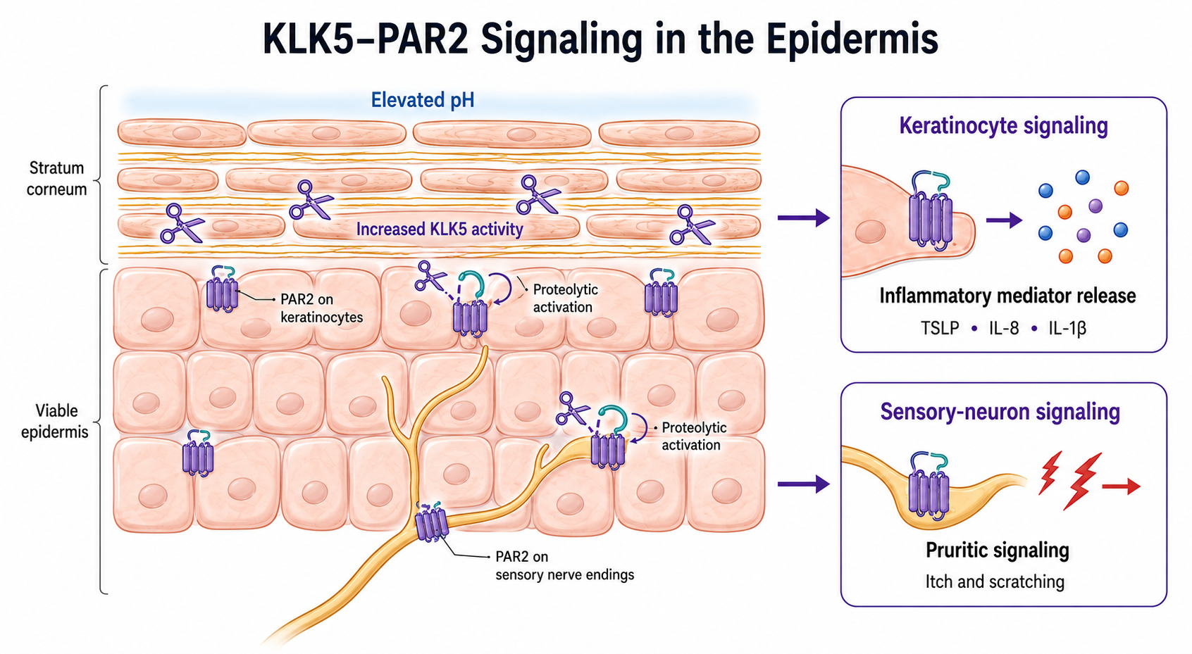
Excess kallikrein activity does more than degrade corneodesmosomes. KLK5 can also activate protease-activated receptor 2 (PAR2), a cell-surface receptor expressed by keratinocytes and cutaneous sensory neurons. Unlike a conventional receptor that is activated when a soluble ligand binds to it, PAR2 is activated when a protease cleaves its extracellular domain. Conditions that increase unrestrained KLK5 activity can therefore increase PAR2 signaling (Briot et al. 2009).
In keratinocytes, PAR2 activation promotes the production of inflammatory and pruritogenic mediators, including thymic stromal lymphopoietin (TSLP), IL-8 (CXCL8), and IL-1β (Briot et al. 2009; Fan et al. 2024). TSLP is an important epithelial alarmin in AD and helps promote the type 2 immune response characteristic of the disease. In sensory neurons, PAR2 links epidermal protease activity to itch and scratching behavior (Steinhoff et al. 2003; Akiyama, Lerner, and Carstens 2015).
Elevated pH can therefore contribute to disease through two interconnected routes: accelerated barrier degradation through altered enzyme kinetics and increased inflammatory and pruritic signaling through PAR2. A murine model of AD demonstrated this connection directly, identifying skin surface pH as a “master switch” governing KLK5-mediated barrier destruction (Jang et al. 2016).
How This Mechanism Contributes to Atopic Dermatitis
Atopic dermatitis is among the best-documented human examples of pH-dependent barrier dysregulation. Clinical studies consistently report higher skin surface pH in AD, with pH often increasing alongside disease activity and severity (Eberlein-König, Schäfer, and Huss 2000; Knor et al. 2011; Proksch 2018; Danby and Cork 2018).
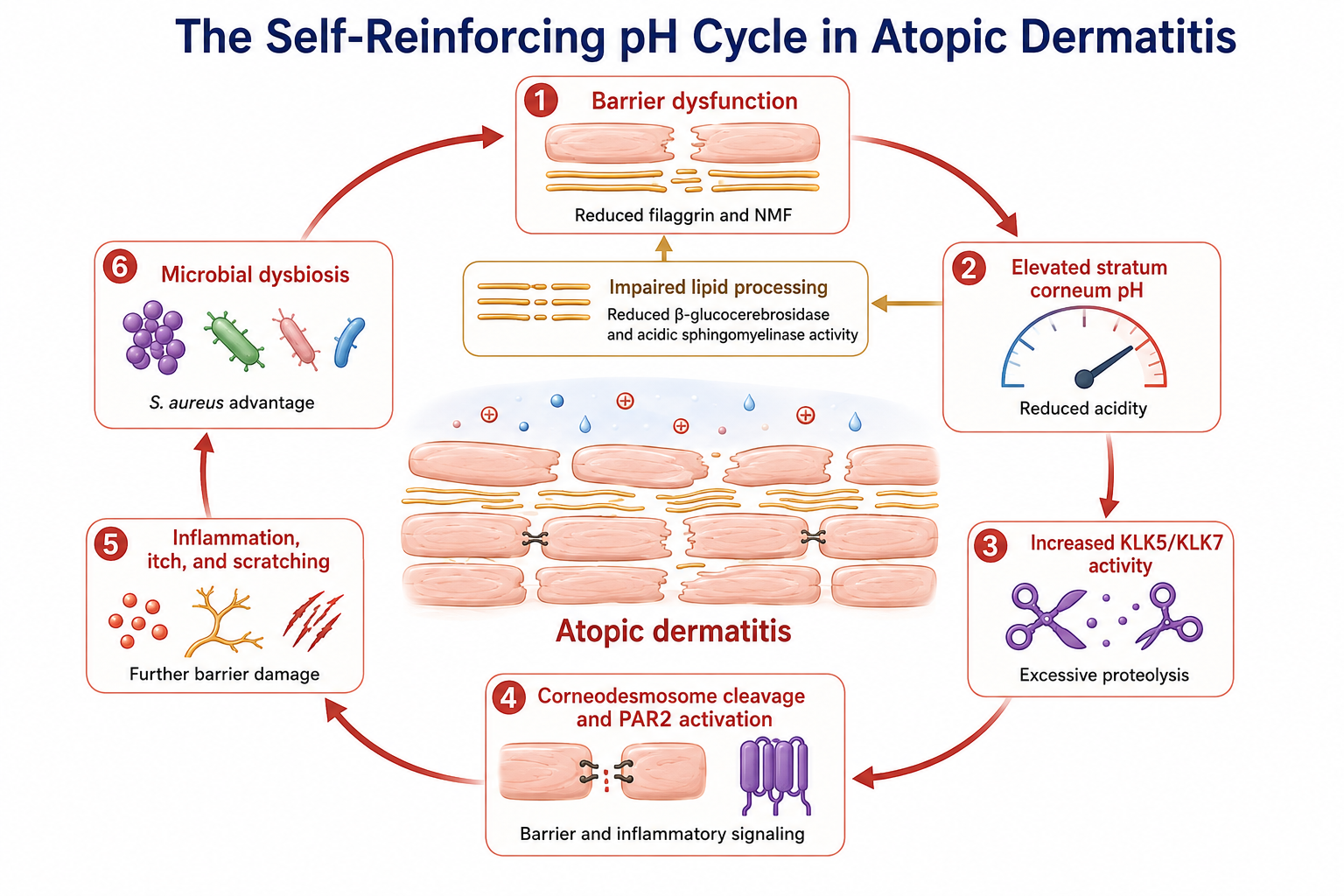
Several features of AD converge to raise pH: reduced filaggrin and natural moisturizing factor, impaired lipid processing, increased protease activity, and microbial dysbiosis. Each of these abnormalities can also be worsened by rising pH, creating a self-reinforcing cycle (Danby and Cork 2018):
Elevated pH increases kallikrein activity, accelerating corneodesmosome degradation and barrier disruption (Hachem et al. 2003, 2005).
Greater KLK5 activity increases PAR2 signaling, promoting inflammation and itch through epithelial mediators and sensory-neuron pathways (Briot et al. 2009; Jang et al. 2016; Steinhoff et al. 2003).
A less acidic surface alters microbial selection. As pH approaches neutrality, Staphylococcus aureus grows more readily and can increase virulence-factor production and biofilm formation, while protective commensals may lose their ecological advantage (Costa and Horswill 2022; Iyer, Raut, and Dasgupta 2021). S. aureus colonization is strongly associated with AD flares.
Together, these processes help explain why AD behaves as a self-sustaining cycle rather than as a single fixed defect. Barrier damage can raise local pH; elevated pH increases protease activity and PAR2 signaling; inflammation, itch, and scratching further damage the barrier; and microbial dysbiosis reinforces the disruption (Danby and Cork 2018).
This pattern is not unique to AD. Elevated pH and related barrier abnormalities have also been described in irritant contact dermatitis, diaper dermatitis, ichthyosis vulgaris, Netherton syndrome, and chronologically aged skin. These conditions begin differently but can converge on disruption of the same pH-dependent regulatory system (Proksch 2018; Stamatas et al. 2011; Gruber et al. 2011; Kilic et al. 2019).
Why a Single pH Measurement Does Not Tell the Whole Story
It may be tempting to reduce this biology to a simple instruction: lower the pH and the problem is solved. The evidence suggests otherwise. Because kallikrein activity responds continuously to the surrounding chemical environment, protease regulation depends not only on the pH reached but also on how long physiological acidity is maintained. A brief change in surface pH does not necessarily restore the deeper buffering and acidification processes of the stratum corneum.
Experimental findings support this distinction. Restoring and maintaining physiological acidity can accelerate barrier recovery, reduce excessive protease activity, and limit inflammatory signaling, whereas transient or superficial acidification may be less reliable (Hachem et al. 2010; Kilic et al. 2019; H.-J. Lee et al. 2015). The relevant biological goal is therefore not a single acidic reading, but the sustained re-establishment of the epidermis’s acid-generating and buffering environment.
The Takeaway
The acid mantle is a regulatory system, not merely a protective film. Under normal mildly acidic conditions, kallikrein proteases remain active enough to support orderly desquamation but sufficiently restrained to preserve barrier cohesion. When pH rises, these enzymes become more active, accelerating corneodesmosome breakdown. KLK5 can also activate PAR2, linking altered pH to inflammatory mediators and itch signaling. At the same time, higher pH can impair lipid-processing enzymes and favor microbial dysbiosis.
In atopic dermatitis, these mechanisms form a reinforcing cycle of elevated pH, barrier disruption, inflammation, itch, scratching, and microbial imbalance. This is why pH is increasingly understood not only as a marker of AD but as a mechanistic contributor to the disease—and why restoring durable physiological acidity requires more than producing a brief change at the surface.
Selected References
Akiyama, Tasuku, Ethan A. Lerner, and E. Carstens. 2015. “Protease-Activated Receptors and Itch.” In Pharmacology of Itch, edited by Alan Cowan and Gil Yosipovitch, vol. 226. Handbook of Experimental Pharmacology. Springer Berlin Heidelberg. https://doi.org/10.1007/978-3-662-44605-8_13
Borgoño, Carla A., Iacovos P. Michael, Nahoko Komatsu, et al. 2007. “A Potential Role for Multiple Tissue Kallikrein Serine Proteases in Epidermal Desquamation.” Journal of Biological Chemistry 282 (6): 3640–52. https://doi.org/10.1074/jbc.M607567200
Brattsand, Maria, Kristina Stefansson, Christine Lundh, Ylva Haasum, and Torbjörn Egelrud. 2005. “A Proteolytic Cascade of Kallikreins in the Stratum Corneum.” Journal of Investigative Dermatology 124 (1): 198–203. https://doi.org/10.1111/j.0022-202X.2004.23547.x
Briot, Anaïs, Céline Deraison, Matthieu Lacroix, et al. 2009. “Kallikrein 5 Induces Atopic Dermatitis–like Lesions through PAR2-Mediated Thymic Stromal Lymphopoietin Expression in Netherton Syndrome.” Journal of Experimental Medicine 206 (5): 1135–47. https://doi.org/10.1084/jem.20082242
Brooks, Sarah G., Rami H. Mahmoud, Rachel R. Lin, Joachim W. Fluhr, and Gil Yosipovitch. 2025. “The Skin Acid Mantle: An Update on Skin pH.” Journal of Investigative Dermatology 145 (3): 509–21. https://doi.org/10.1016/j.jid.2024.07.009
Caubet, Cécile, Nathalie Jonca, Maria Brattsand, et al. 2004. “Degradation of Corneodesmosome Proteins by Two Serine Proteases of the Kallikrein Family, SCTE/KLK5/hK5 and SCCE/KLK7/hK7.” Journal of Investigative Dermatology 122 (5): 1235–44. https://doi.org/10.1111/j.0022-202X.2004.22512.x
Costa, Flavia G., and Alexander R. Horswill. 2022. “Overcoming pH Defenses on the Skin to Establish Infections.” PLOS Pathogens 18 (5): e1010512. https://doi.org/10.1371/journal.ppat.1010512
Danby, Simon G., and Michael J. Cork. 2018. “pH in Atopic Dermatitis.” In Current Problems in Dermatology, edited by Christian Surber, Christoph Abels, and Howard Maibach, vol. 54. S. Karger AG. https://doi.org/10.1159/000489523
Eberlein-König, B., T. Schäfer, and J. Huss. 2000. “Skin Surface pH, Stratum Corneum Hydration, Trans-Epidermal Water Loss and Skin Roughness Related to Atopic Eczema and Skin Dryness in a Population of Primary School Children: Clinical Report.” Acta Dermato-Venereologica 80 (3): 188–91. https://doi.org/10.1080/000155500750042943
Elias, Peter M. 2015. “Stratum Corneum Acidification: How and Why?” Experimental Dermatology 24 (3): 179–80. https://doi.org/10.1111/exd.12596
Fan, Mengjie, Xiaoyao Fan, Yangfan Lai, et al. 2024. “Protease-Activated Receptor 2 in Inflammatory Skin Disease: Current Evidence and Future Perspectives.” Frontiers in Immunology 15 (September): 1448952. https://doi.org/10.3389/fimmu.2024.1448952
Fluhr, Joachim W., and Peter M. Elias. 2002. “Stratum Corneum pH: Formation and Function of the ‘Acid Mantle.’” Exogenous Dermatology 1 (4): 163–75. https://doi.org/10.1159/000066140
Gruber, Robert, Peter M. Elias, Debra Crumrine, et al. 2011. “Filaggrin Genotype in Ichthyosis Vulgaris Predicts Abnormalities in Epidermal Structure and Function.” The American Journal of Pathology 178 (5): 2252–63. https://doi.org/10.1016/j.ajpath.2011.01.053
Hachem, Jean-Pierre, Debra Crumrine, Joachim Fluhr, Barbara E. Brown, Kenneth R. Feingold, and Peter M. Elias. 2003. “pH Directly Regulates Epidermal Permeability Barrier Homeostasis, and Stratum Corneum Integrity/Cohesion.” Journal of Investigative Dermatology 121 (2): 345–53. https://doi.org/10.1046/j.1523-1747.2003.12365.x
Hachem, Jean-Pierre, Mao-Quiang Man, Debra Crumrine, et al. 2005. “Sustained Serine Proteases Activity by Prolonged Increase in pH Leads to Degradation of Lipid Processing Enzymes and Profound Alterations of Barrier Function and Stratum Corneum Integrity.” Journal of Investigative Dermatology 125 (3): 510–20. https://doi.org/10.1111/j.0022-202X.2005.23838.x
Hachem, Jean-Pierre, Truus Roelandt, Nanna Schürer, et al. 2010. “Acute Acidification of Stratum Corneum Membrane Domains Using Polyhydroxyl Acids Improves Lipid Processing and Inhibits Degradation of Corneodesmosomes.” Journal of Investigative Dermatology 130 (2): 500–510. https://doi.org/10.1038/jid.2009.249
Iyer, Vidula, Janhavi Raut, and Anindya Dasgupta. 2021. “Impact of pH on Growth of Staphylococcus Epidermidis and Staphylococcus Aureus in Vitro.” Journal of Medical Microbiology 70 (9). https://doi.org/10.1099/jmm.0.001421
Jang, Hyosun, Akira Matsuda, Kyungsook Jung, et al. 2016. “Skin pH Is the Master Switch of Kallikrein 5-Mediated Skin Barrier Destruction in a Murine Atopic Dermatitis Model.” Journal of Investigative Dermatology 136 (1): 127–35. https://doi.org/10.1038/JID.2015.363
Kilic, Ana, Clarissa Masur, Hubert Reich, et al. 2019. “Skin Acidification with a Water-in-Oil Emulsion (pH 4) Restores Disrupted Epidermal Barrier and Improves Structure of Lipid Lamellae in the Elderly.” The Journal of Dermatology 46 (6): 457–65. https://doi.org/10.1111/1346-8138.14891
Knor, Tanja, Ajša Meholjić-Fetahović, and Aida Mehmedagić. 2011. “Stratum Corneum Hydration and Skin Surface pH in Patients with Atopic Dermatitis.” Acta Dermatovenerologica Croatica 19 (4): 242–47.
Lee, Hae-Jin, Noo Ri Lee, Minyoung Jung, Dong Hye Kim, and Eung Ho Choi. 2015. “Atopic March from Atopic Dermatitis to Asthma-Like Lesions in NC/Nga Mice Is Accelerated or Aggravated by Neutralization of Stratum Corneum but Partially Inhibited by Acidification.” Journal of Investigative Dermatology 135 (12): 3025–33. https://doi.org/10.1038/jid.2015.333
Proksch, Ehrhardt. 2018. “pH in Nature, Humans and Skin.” The Journal of Dermatology 45 (9): 1044–52. https://doi.org/10.1111/1346-8138.14489
Schade, H., and A. Marchionini. 1928. “Der Säuremantel der Haut (Nach Gaskettenmessungen).” Klinische Wochenschrift 7 (1): 12–14. https://doi.org/10.1007/BF01711684
Stamatas, Georgios N., Charles Zerweck, Gary Grove, and Katharine M. Martin. 2011. “Documentation of Impaired Epidermal Barrier in Mild and Moderate Diaper Dermatitis In Vivo Using Noninvasive Methods.” Pediatric Dermatology 28 (2): 99–107. https://doi.org/10.1111/j.1525-1470.2011.01308.x
Steinhoff, Martin, Ulrich Neisius, Akihiko Ikoma, et al. 2003. “Proteinase-Activated Receptor-2 Mediates Itch: A Novel Pathway for Pruritus in Human Skin.” The Journal of Neuroscience 23 (15): 6176–80. https://doi.org/10.1523/JNEUROSCI.23-15-06176.2003
Surber, Christian, Philippe Humbert, Christoph Abels, and Howard Maibach. 2018. “The Acid Mantle: A Myth or an Essential Part of Skin Health?” In Current Problems in Dermatology, edited by Christian Surber, Christoph Abels, and Howard Maibach, vol. 54. S. Karger AG. https://doi.org/10.1159/000489512
Takagi, Yutaka, Ernst Kriehuber, Genji Imokawa, Peter M. Elias, and Walter M. Holleran. 1999. “β-Glucocerebrosidase Activity in Mammalian Stratum Corneum.” Journal of Lipid Research 40 (5): 861–69. https://doi.org/10.1016/S0022-2275(20)32121-0